Models of Neonatal Hyperoxgenation: Bronchopulmonary Dysplasia and Oxygen-Induced Retinopathy
Neonatal hyperoxygenation has been demonstrated to produce at least two significant syndromes, Bronchopulmonary Dysplasia and Retinopathy of Prematurity in children. Developing treatments for these two syndromes represents an unmet medical need. Here at CBI, we have developed and validated robust models for both of these clinical indications for the purposes of assessing mechanism of action and for assessing the activity of treatment modalities. The preclinical models employ neonatal murine species. Placing pups in a high oxygen environment for 2 weeks induces both bronchopulmonary dysplasia and oxygen-induced retinopathy that are translationally useful and robust models of the human disease.
Hyperoxygenation Models at CBI
- Bronchopulmonary Dysplasia- For more information, visit our pulmonary white paper or download our presentation.
- Oxygen-induced retinopathy (retinopathy of prematurity)- For more information, visit our OIR white paper or download our presentation.
- Angiotensin-II induced retinal vascular leakage
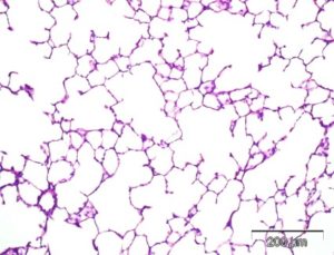
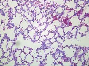
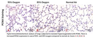
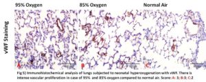
CBI’s in-house Histology lab and expertise in Immunohistochemistry provide validated, superior methods of assessment including our proprietary Phase Contrast Analysis methodology in comparison to standard MLI assessments of alveolar walls.
IHC assessments may include:
- Collagen type 1
- SMA 1
- CD68
- vWf
- CD8
- PCNA
- KI67
Click here to learn more about our validated method
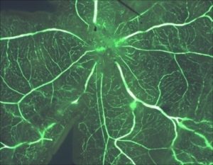
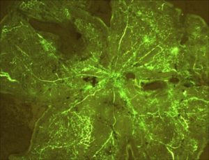
Exposure of neonatal infant pups to 2 weeks of hyper-oxygenation followed by return to room air leads to a proliferative neovascularization of the retina that models the Retinopathy of Prematurity in premature infants. CBI offers a sophisticated and validated model of oxygen-induced retinopathy in neonatal murine species.


