Robert Sukhu, Qin Zhang, Isaac Hakim, Ali Ebramhimnejad, Carol Meschter, Michelle Kelley
Comparative Biosciences, Inc., Sunnyvale, CA 94085
Click here for a downloadable version
Thermal and UV burns represent an unmet medical need, particularly in defense or military settings. CBI has a number of humane, validated and optimized superficial, partial thickness and full thickness burn models in rodents and pigs, including mini and Duroc pigs. We have evaluated a number of therapeutic regimes including biologics and stem cell treatments as well as small molecules and immunomodulatory agents.
Thermal Burn Modeling in Göttingen Minipigs
We at CBI are very pleased to be asked to prepare an article on our thermal burn modeling in Göttingen Pigs for Marshall Farms. We have been using Marshall Farms, Inc. pigs and dogs at our facilities for over 15 years for a wide variety of toxicology, pharmacokinetic, and pharmacology studies. A burn is a tissue injury resulting from exposure to various noxious agents. For purposes of this article, we will limit the discussion to burns of the skin resulting from heat. Burn therapy has become a research priority, in part due to the recent combat experience and concern over the possibility of massive terrorist attacks. Consequently, our clients have become interested in investigating various treatments and methods in this therapeutic area. At CBI we have developed some excellent models of burns and other dermal wounds using Göttingen minipigs. We adapted models published by Singer et al. (2011) and Branski et al. (2008) to our own expertise and requirements as well as the needs of our clients. As a contract research house, our mission is not so much to explore the nature of burn tissue injury or mechanisms of healing, but to quickly and thoroughly evaluate the safety and efficacy of new treatments, in way that would meet regulatory standards. Our models are effective, reproducible and humane. We have also used Red Duroc pigs and other strains of pigs successfully.
Types of burns
There are 3 major types of dermal thermal burns:
- First Degree or Superficial: Superficial epidermis with hyperemia and no blistering
- Second or Partial Thickness: Affects epidermis and dermis-superficial (epidermis and papillary dermis) or deep (reticular dermis) (see Fig. 1)
- Third or Full thickness: Affects epidermis and dermis and extends to the subcutaneous tissue (see Fig. 2)
- Some authorities also describe Fourth Degree burns, but these typically require extensive surgical treatment, including amputation, and are beyond the scope of this article.
Some authorities also describe Fourth Degree burns, but these typically require extensive surgical treatment, including amputation, and are beyond the scope of this article.

Pain Management
Pain is always a concern in wound studies and must be managed carefully. We use intraoperative pre-emptive pain relief followed by a fentanyl patch with supplemental buprenorphine or butorphanol. Pigs are carefully monitored throughout the study for signs of pain or distress. CBI had an unannounced USDA inspection while we were performing one of these models and they found no deficiencies. It is worth noting that in humans, first- and superficial second-degree burns are reported to be more painful than deep second- and third-degree burns.
Anesthesia and Surgery
For anesthesia, we use a combination of pre-emptive pain relief, fluids, sedation induction, intubation and isoflurane maintenance. EKG, body temperature, blood pressure, respiratory rate, hemoglobin saturation are monitored. Animals are on heating pads. Strict asepsis is critical to all the surgical procedures.
Burn Induction
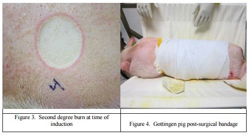
We have developed a method to consistently induce a uniform burn that is either a first-, second-(superficial or deep) or third-degree burn (See Fig. 3).
Bandage changing and dressings
Burn and wound management generally requires some sort of wound covering (see Fig. 4). The individual study protocol generally specifies the type of bandage changing and dressings. We are careful to keep stress and excitement and pain to a minimum during this sort of handling. The preconditioning of the animal is helpful in this regard.
Burn treatment modalities
There is a variety of treatment modalities that are of current interest including:
- Topical or systemic administration
- Escharotomy and debridement
- Stem cell therapies
- Autologous cells and stem cells
- Small molecules
- Biologics and immunomodulatory agents
- Bandaging or wound dressings
- Sealants
- Minced preps, split and full thickness grafting
Each modality may require variations on the model. Each study protocol is custom tailored to the needs of the client.
Additional Surgical Procedures and Biopsies
Protocols often call for additional procedures such as escharectomy, skin grafts or biopsies at intervals following the original burn procedure. With these procedures, pigs are susceptible to problems with anesthesia and recovery. Consequently, we have developed some techniques and methods to optimize these secondary procedures.
Thermal Burn Modeling Endpoints
As part of a program addressing the three Rs, we feel it is very important to obtain as much information from each animal as possible. At minimum we assess the following:
- Wound healing progression with digital image analysis of wound size and closure
- Draize scoring
- Transepidermal water loss
- Body temperature and body weights, food consumption
- Hematology and clinical chemistry
- Serum and tissue markers of inflammation
- Serial biopsies
- Histopathology and photomicroscopy
- Special stains and immunohistochemistry
- Histomorphometry
- Custom upon request
- Complete, timely reporting suitable for regulatory submission (GLP and non-GLP studies)
Preconditioning
For preconditioning, pigs receive a physical exam and routine blood work. Pigs are acclimated to the facility and enrichment program. They are also handled, and practiced being carried and putting into a sling with positive re-enforcement. This helps facilitate bandage changing, scoring of wounds, biopsies and photography. It is important in burn and wound studies that the pigs are kept in the most stress free environment possible so that there is a little interference with the healing process as possible.
Nutrition
High quality nutrition and good food consumption is important to success of the study. We provide an optimized diet plus supplements such as fruit and alfalfa.
Environmental Enrichment
Environmental enrichment is an important aspect of managing pigs on a wound study. They need to be kept comfortable and occupied to reduce fretting and damaging their bandages. Radios, television, toys, fruit, hay, alfalfa cubes, exercise pens, petting and scratching, are helpful.
Why we like Göttingens at CBI
As we optimized this model, we worked with several strains of pigs and settled on the Marshall Farms Göttingen Minipig. The pigs are physically uniform, free of disease, gentle, have good appetites and easy to handle and carry. Further, they handle multiple anesthesias, multiple sedations and restraint well. Subclinical conditions, particularly respiratory infections, can lead to poor surgical and anesthesia outcomes in swine, but the Göttingens are free of pulmonary pathogens. Another important feature of these pigs is their uniformity in size and shape and their growth curves. This is important in following wound progression and healing over time. We can also conduct modeling in disease states such as hypertension, diabetes and immune suppression in burn- and wound-healing studies to mimic human disorders.
For our burn studies, we design our studies to optimize the success of the study. This means that we have well designed studies with adequate numbers of animals and study endpoints to assure relevance of the data. Initially, preconditioning, acclimation and environmental enrichment are key parts of the study design. this is followed by the procedure and treatment, as well as recovery and healing prior to necropsy.
Summary
In summary, we at CBI have developed and optimized a 1st, 2nd and 3rd degree thermal dermal burn model using the Gottingen minipig. For our burn studies, we design our studies to optimize the success of the study. This leads to well designed studies with adequate numbers of animals and study endpoints to assure relevance of the data. Initially, preconditioning, acclimation and environmental enrichment are key parts of the study design. This is followed by the procedure and treatment, as well as recovery and healing prior to necropsy. Our methods allow us to evaluate various treatment modalities to assess healing in a manner that is both useful and humane.
References
Singer AJ, Hirth D, McClain SA, Crawford L, Lin F, Clark RA. Validation of a vertical progression porcine burn model. J Burn Care Res. 2011 Nov-Dec; 32(6):638-46.
Branski LK, Mittermayr R, Herndon DN, Norbury WB, Masters OE, Hofmann M, Traber DL, Redl H, Jeschke MG. A porcine model of full-thickness burn, excision and skin autografting. Burns. 2008 Dec; 34(8):1119-27.
FoubersP, BarillasS, GonzalezAD, AlfonsoZ, ZhaoS, HakimI, MeschterC, TenenhausM, FraserJK. Uncultured adiposederived regenerative cells (ADRCs) seeded in collagen scaffold improves dermal regeneration, enhancing early vascularization and structural organization following thermal burns. Burns. Nov;41(7):1504-16, 2015.